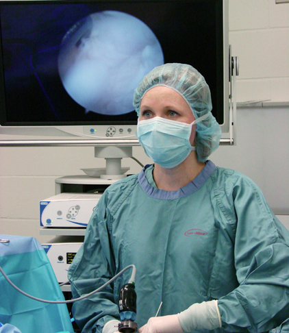
"Right now, there's no peer-reviewed research that actually shows they work even though there are a lot of claims that they do," says Dr. Rhea Plesman, a small animal surgery resident at the Western College of Veterinary Medicine (WCVM).
Along with her resident advisor, WCVM small animal surgeon Dr. Kathleen Linn, Plesman planned to investigate the effectiveness of the knee brace in dogs for her graduate research project.
But after reviewing previous research, Plesman realized that she first needed to develop a standardized, non-invasive method of evaluating the knee (or stifle) joint following rupture of the cranial cruciate ligament.
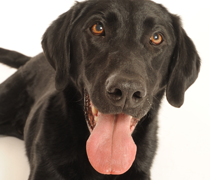
The most common orthopedic condition seen in veterinary medicine, rupture of the CCL in canine patients is similar to an ACL (anterior cruciate ligament) tear in people. The injury can occur at any age and in any breed or size of dog with Labrador retrievers being one of the more commonly affected breeds.
The CCL plays a key role in stabilizing a dog's stifle joint, and a rupture often presents as a degenerative process in dogs.
"Owners often observe a waxing and waning type of lameness," explains Plesman. "An affected dog will appear sore, improve for a time and then become sore again until the entire ligament completely ruptures."
After a complete tear, an immediate, non-weight bearing lameness can occur that may improve over time. Owners may notice their dog no longer sits squarely and may sit with their legs off to the side. If the ligaments of both knees are damaged, affected dogs may have trouble going up and down stairs and difficulty rising from a sitting position.
Some patients may be medically managed with anti-inflammatory medications, exercise restriction and joint supplements. But most require surgical intervention followed by physical rehabilitation in order to regain as much function of the stifle as possible.
Repeat radiographic techniques
When the CCL ruptures, the tibia moves forward in relation to the femur creating instability — a condition called cranial tibial subluxation (CTS).
"The current method of measuring CTS involves surgically implanting metal markers directly into the femur and tibia allowing for easy visualization on an X-ray," says Plesman. "But this isn't an ideal procedure for the live patient."
There are several physical exam findings that also help to diagnose CCL rupture: both the cranial drawer test and the cranial tibial thrust test assess the amount of forward movement of the tibia in relation to the femur. As well, the presence of thickened fibrous tissue surrounding the knee joint and fluid build-up within the joint capsule may indicate ligament rupture.
Radiographs are a helpful diagnostic tool, but Plesman found current techniques and measurements to be poorly defined and not standardized. With her study, the small animal surgery resident aimed to develop a repeatable radiographic technique for assessing CTS in clinically affected dogs.
With funds provided by the Canadian Kennel Club (CKC) and the veterinary college's Companion Animal Health Fund (CAHF), Plesman analyzed 10 randomly selected clinical cases of cranial cruciate ligament rupture that presented to the WCVM's Veterinary Medical Centre between 2010 and 2011.
With the help of Dr. Ajay Sharma, a board-certified radiologist and former WCVM associate professor of medical imaging, Plesman identified a series of radiographic landmarks on the femoral and tibial bones that showed potential to serve as points for measuring CTS.
"We chose landmarks that were easily seen on an X-ray," she says. "We also chose points that wouldn't be obscured by arthritic changes as well as points of reference commonly used for guiding surgical repair."
In the end, the research team chose six anatomical landmarks: two near the end of the femur and four at the top of the tibia.
Over the span of a year, Plesman collected 20 normal hind limbs from medium to large breed canine cadavers. With all of the tissues above the stifle joint cleared away, the limb was inserted into a custom-made limb press testing apparatus. To mimic normal weight bearing, a load equal to 20 per cent of the dog's body weight was applied to the leg using this apparatus.
For each specimen, radiographs were taken before and after cutting the cranial cruciate ligament, with and without the metal bone markers in place.
Plesman, Sharma and Dr. Peter Gilbert, a WCVM associate professor of small animal surgery, examined the radiographs and measured each landmark in relation to each other.
Measurements with the ligament intact were compared to measurements with the ligament cut to determine how much the tibia moved forward. As well, comparisons were made between the selected anatomical landmarks versus the metal bone markers.
The best of the bone markers
After assessing variability, the team concluded that the measurement of CTS from the caudal limit of the intercondylar fossa (a deep notch at the end of the back of the femur) to the intercondylar eminence (a raised ridge at the top of the tibia) was the most reliable. Both of these bony landmarks are near the attachment site for the cranial cruciate ligament.
Unexpectedly, the researchers observed a poor correlation between the anatomic landmarks and the bone markers.
"It's unusual since the use of these bone markers is considered the current gold standard for CTS measurement," says Plesman. She adds that this may have occurred for any number of reasons including the effects of internal rotation of the knee when the cranial cruciate ligament is ruptured.
The two-year study's results offer an improved method of radiographically diagnosing cruciate rupture. But more importantly, it significantly advances veterinary research related to cruciate disease.
"It offers some building blocks for future clinical studies," says Plesman. "It will also help to better evaluate the methods we're using to treat cruciate ruptures. We can assess the stifle post-operatively and figure out if there's something we could be doing better."
With this preliminary work behind them, the research team has begun the next phase in collaboration with Dr. James Johnston, an assistant professor in the University of Saskatchewan's College of Engineering. Johnston will play an important role in the design and testing of a tibial compression device that the research team hopes to use on a live dog.
The device will mimic the cranial tibial thrust test that veterinarians currently perform manually, essentially standardizing it so the same pressure and technique is used every time.
"We also want to develop a method of taking standing radiographs of the stifle," says Plesman.
With the knowledge gained from these investigations, the team can then start to look more intensively at the effectiveness of supportive stifle braces for dogs with cruciate ligament injuries.
"We hope to assess affected dogs using the tibial compression device before and after applying the brace," says Plesman. "It will be interesting to see if the brace provides stability and eliminates tibial thrust as many marketers claim."
Robyn Thrasher of Edmonton, Alta., is a third-year veterinary student at the WCVM. Robyn is a WCVM research communications intern as well as a summer student in the WCVM Veterinary Medical Centre during the summer of 2012.
Along with her resident advisor, WCVM small animal surgeon Dr. Kathleen Linn, Plesman planned to investigate the effectiveness of the knee brace in dogs for her graduate research project.
But after reviewing previous research, Plesman realized that she first needed to develop a standardized, non-invasive method of evaluating the knee (or stifle) joint following rupture of the cranial cruciate ligament.
The most common orthopedic condition seen in veterinary medicine, rupture of the CCL in canine patients is similar to an ACL (anterior cruciate ligament) tear in people. The injury can occur at any age and in any breed or size of dog with Labrador retrievers being one of the more commonly affected breeds.
The CCL plays a key role in stabilizing a dog's stifle joint, and a rupture often presents as a degenerative process in dogs.
"Owners often observe a waxing and waning type of lameness," explains Plesman. "An affected dog will appear sore, improve for a time and then become sore again until the entire ligament completely ruptures."
After a complete tear, an immediate, non-weight bearing lameness can occur that may improve over time. Owners may notice their dog no longer sits squarely and may sit with their legs off to the side. If the ligaments of both knees are damaged, affected dogs may have trouble going up and down stairs and difficulty rising from a sitting position.
Some patients may be medically managed with anti-inflammatory medications, exercise restriction and joint supplements. But most require surgical intervention followed by physical rehabilitation in order to regain as much function of the stifle as possible.
Repeat radiographic techniques
When the CCL ruptures, the tibia moves forward in relation to the femur creating instability — a condition called cranial tibial subluxation (CTS).
"The current method of measuring CTS involves surgically implanting metal markers directly into the femur and tibia allowing for easy visualization on an X-ray," says Plesman. "But this isn't an ideal procedure for the live patient."
There are several physical exam findings that also help to diagnose CCL rupture: both the cranial drawer test and the cranial tibial thrust test assess the amount of forward movement of the tibia in relation to the femur. As well, the presence of thickened fibrous tissue surrounding the knee joint and fluid build-up within the joint capsule may indicate ligament rupture.
Radiographs are a helpful diagnostic tool, but Plesman found current techniques and measurements to be poorly defined and not standardized. With her study, the small animal surgery resident aimed to develop a repeatable radiographic technique for assessing CTS in clinically affected dogs.
With funds provided by the Canadian Kennel Club (CKC) and the veterinary college's Companion Animal Health Fund (CAHF), Plesman analyzed 10 randomly selected clinical cases of cranial cruciate ligament rupture that presented to the WCVM's Veterinary Medical Centre between 2010 and 2011.
With the help of Dr. Ajay Sharma, a board-certified radiologist and former WCVM associate professor of medical imaging, Plesman identified a series of radiographic landmarks on the femoral and tibial bones that showed potential to serve as points for measuring CTS.
"We chose landmarks that were easily seen on an X-ray," she says. "We also chose points that wouldn't be obscured by arthritic changes as well as points of reference commonly used for guiding surgical repair."
In the end, the research team chose six anatomical landmarks: two near the end of the femur and four at the top of the tibia.
Over the span of a year, Plesman collected 20 normal hind limbs from medium to large breed canine cadavers. With all of the tissues above the stifle joint cleared away, the limb was inserted into a custom-made limb press testing apparatus. To mimic normal weight bearing, a load equal to 20 per cent of the dog's body weight was applied to the leg using this apparatus.
For each specimen, radiographs were taken before and after cutting the cranial cruciate ligament, with and without the metal bone markers in place.
Plesman, Sharma and Dr. Peter Gilbert, a WCVM associate professor of small animal surgery, examined the radiographs and measured each landmark in relation to each other.
Measurements with the ligament intact were compared to measurements with the ligament cut to determine how much the tibia moved forward. As well, comparisons were made between the selected anatomical landmarks versus the metal bone markers.
The best of the bone markers
After assessing variability, the team concluded that the measurement of CTS from the caudal limit of the intercondylar fossa (a deep notch at the end of the back of the femur) to the intercondylar eminence (a raised ridge at the top of the tibia) was the most reliable. Both of these bony landmarks are near the attachment site for the cranial cruciate ligament.
Unexpectedly, the researchers observed a poor correlation between the anatomic landmarks and the bone markers.
"It's unusual since the use of these bone markers is considered the current gold standard for CTS measurement," says Plesman. She adds that this may have occurred for any number of reasons including the effects of internal rotation of the knee when the cranial cruciate ligament is ruptured.
The two-year study's results offer an improved method of radiographically diagnosing cruciate rupture. But more importantly, it significantly advances veterinary research related to cruciate disease.
"It offers some building blocks for future clinical studies," says Plesman. "It will also help to better evaluate the methods we're using to treat cruciate ruptures. We can assess the stifle post-operatively and figure out if there's something we could be doing better."
With this preliminary work behind them, the research team has begun the next phase in collaboration with Dr. James Johnston, an assistant professor in the University of Saskatchewan's College of Engineering. Johnston will play an important role in the design and testing of a tibial compression device that the research team hopes to use on a live dog.
The device will mimic the cranial tibial thrust test that veterinarians currently perform manually, essentially standardizing it so the same pressure and technique is used every time.
"We also want to develop a method of taking standing radiographs of the stifle," says Plesman.
With the knowledge gained from these investigations, the team can then start to look more intensively at the effectiveness of supportive stifle braces for dogs with cruciate ligament injuries.
"We hope to assess affected dogs using the tibial compression device before and after applying the brace," says Plesman. "It will be interesting to see if the brace provides stability and eliminates tibial thrust as many marketers claim."
Robyn Thrasher of Edmonton, Alta., is a third-year veterinary student at the WCVM. Robyn is a WCVM research communications intern as well as a summer student in the WCVM Veterinary Medical Centre during the summer of 2012.
Share this story