
A new way to learn about neuroscience
Two professors from the Western College of Veterinary Medicine (WCVM) are developing new teaching methods to simplify the daunting subject of neuroscience for veterinary students.
By Melissa Cavanagh
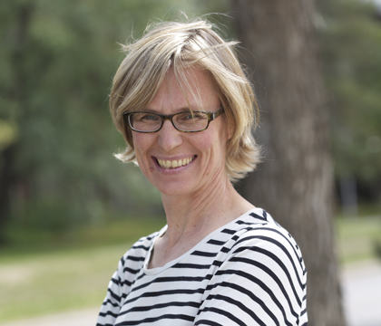
"We want to develop a website to assist with teaching neuroscience throughout the [veterinary] curriculum," says Dr. Gillian Muir, a professor of neuroscience at the WCVM. Muir is working with Dr. Susan Taylor, a professor in the WCVM's Department of Small Animal Clinical Sciences.
"The website won't be text-based learning," explains Muir. "I want [the students] to be able to see it."
Throughout her teaching career, Muir has seen many students struggle with the subject of neuroscience. The most common complaint is that students are unable to visualize the anatomy of the nervous system by looking at a two-dimensional drawing.
"My first thought was to write a textbook . . . but I wanted really strong visual images, and you can't get a 3-D view from a book."
Muir was developing initial plans for a neuroscience website for her students when she discovered that her WCVM colleague was working on a similar project.
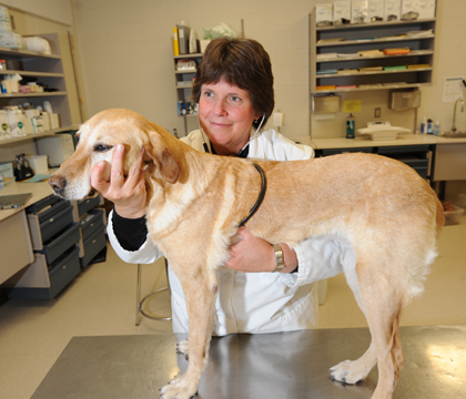
Taylor had plans to organize her library of neuroscience cases during her year-long sabbatical leave. A specialist in small animal internal medicine, Taylor has nearly 350 videos of animals with neurologic lesions as well as a collection of X-rays, MRI and CT studies, lab tests, muscle biopsies and other diagnostic test results that accompany the videos.
Her goal is to have all of the videos accessible in an easy-to-use format: "They don't do anybody any good when they sit in all these boxes on my shelf."
Taylor contacted Muir to see if she would be interested in helping put the cases together and to come up with anatomic descriptions of the location of the neurologic lesion for each case.
"[Gillian] emailed me back right away and said, ‘No way, that's what I'm doing for my sabbatical!'" says Taylor.
Muir is working to develop anatomic models of the nervous system to illustrate the location of pathways and structures important for identifying lesions.. In the meantime, Taylor is working on the videos and cases to accompany the anatomic models. The website will explain how a lesion in a specific area results in abnormal neurologic findings in an animal.
Muir and Taylor are working with eMAP (Media Access and Production) at the U of S to develop a neuroscience website that will be available to WCVM students by next year.
The initial plan is to have WCVM students use the site and review it so the web development team can make some minor tweaks if necessary. Once the site is ready, Muir and Taylor eventually want to make it available to students and instructors at other veterinary colleges.
"Our hope is that it will be of interest to practitioners as well. But our real audience is the students," says Taylor.
She adds that the courses won't be taught off the website. Instead, the site will act as an additional tool that students can access after class to gain a better visual understanding of the topic.
"Watching videos of dogs and cats with lesions affecting specific regions of the brain or spinal cord will teach students to recognize the gait associated with lesions in each location, building confidence in their approach to neurologic patients," Taylor explains.
"A first-year student might click on anatomic localization . . . and they would be able to see 10 or 15 videos of dogs that had lesions [in a specific area]. They might not even go through the cases in terms of looking at all the diagnostic results," explains Taylor.
On the other hand, a third- year student may be more apt to approach the site from a case point of view. "They could temporarily link back to the anatomy [section] to remind themselves what tracts are there and why they're seeing those particular neurologic signs. So I think everyone will use the site, but they'll use it in different ways."
Taylor, who is staying in Saskatoon for most of her sabbatical leave, will continue to teach clinical neurology lectures and labs to second and third year students at the WCVM as she works on the website project. She will also spend some time with veterinary neurologists at North Carolina State University and the University of Tennessee, seeing cases and collecting videos for the project.
Muir is working in a much more exotic locale: she's spending her 12-month sabbatical leave in France with her husband and three young daughters. For more details, visit her family's two blogs: freakytink.com (written by Muir and her husband) and 3kittiesinfrance.blogspot.com (written by Muir's daughters).
Melissa Cavanagh of Winnipeg, Man., is a second-year veterinary student and was the WCVM's research communications intern for the summer of 2013.
"The website won't be text-based learning," explains Muir. "I want [the students] to be able to see it."
Throughout her teaching career, Muir has seen many students struggle with the subject of neuroscience. The most common complaint is that students are unable to visualize the anatomy of the nervous system by looking at a two-dimensional drawing.
"My first thought was to write a textbook . . . but I wanted really strong visual images, and you can't get a 3-D view from a book."
Muir was developing initial plans for a neuroscience website for her students when she discovered that her WCVM colleague was working on a similar project.
Taylor had plans to organize her library of neuroscience cases during her year-long sabbatical leave. A specialist in small animal internal medicine, Taylor has nearly 350 videos of animals with neurologic lesions as well as a collection of X-rays, MRI and CT studies, lab tests, muscle biopsies and other diagnostic test results that accompany the videos.
Her goal is to have all of the videos accessible in an easy-to-use format: "They don't do anybody any good when they sit in all these boxes on my shelf."
Taylor contacted Muir to see if she would be interested in helping put the cases together and to come up with anatomic descriptions of the location of the neurologic lesion for each case.
"[Gillian] emailed me back right away and said, ‘No way, that's what I'm doing for my sabbatical!'" says Taylor.
Muir is working to develop anatomic models of the nervous system to illustrate the location of pathways and structures important for identifying lesions.. In the meantime, Taylor is working on the videos and cases to accompany the anatomic models. The website will explain how a lesion in a specific area results in abnormal neurologic findings in an animal.
Muir and Taylor are working with eMAP (Media Access and Production) at the U of S to develop a neuroscience website that will be available to WCVM students by next year.
The initial plan is to have WCVM students use the site and review it so the web development team can make some minor tweaks if necessary. Once the site is ready, Muir and Taylor eventually want to make it available to students and instructors at other veterinary colleges.
"Our hope is that it will be of interest to practitioners as well. But our real audience is the students," says Taylor.
She adds that the courses won't be taught off the website. Instead, the site will act as an additional tool that students can access after class to gain a better visual understanding of the topic.
"Watching videos of dogs and cats with lesions affecting specific regions of the brain or spinal cord will teach students to recognize the gait associated with lesions in each location, building confidence in their approach to neurologic patients," Taylor explains.
"A first-year student might click on anatomic localization . . . and they would be able to see 10 or 15 videos of dogs that had lesions [in a specific area]. They might not even go through the cases in terms of looking at all the diagnostic results," explains Taylor.
On the other hand, a third- year student may be more apt to approach the site from a case point of view. "They could temporarily link back to the anatomy [section] to remind themselves what tracts are there and why they're seeing those particular neurologic signs. So I think everyone will use the site, but they'll use it in different ways."
Taylor, who is staying in Saskatoon for most of her sabbatical leave, will continue to teach clinical neurology lectures and labs to second and third year students at the WCVM as she works on the website project. She will also spend some time with veterinary neurologists at North Carolina State University and the University of Tennessee, seeing cases and collecting videos for the project.
Muir is working in a much more exotic locale: she's spending her 12-month sabbatical leave in France with her husband and three young daughters. For more details, visit her family's two blogs: freakytink.com (written by Muir and her husband) and 3kittiesinfrance.blogspot.com (written by Muir's daughters).
Melissa Cavanagh of Winnipeg, Man., is a second-year veterinary student and was the WCVM's research communications intern for the summer of 2013.