Medical imaging experts brainstorm at U of S
Medical imaging experts from around the globe gathered at the University of Saskatchewan on May 16 to discuss and develop new ways to improve health care for people and animals.
By Melissa Cavanagh"Frontiers in Hybrid Medical Imaging Technology" was a day-long brainstorming workshop that was organized by the Western College of Veterinary Medicine (WCVM), U of S College of Medicine and the Sylvia Fedoruk Canadian Centre for Nuclear Innovation. The workshop's 50-plus participants came from a variety of backgrounds including veterinary medicine, human health care, physics and industry.
Medical imaging has traditionally been used to diagnose illnesses such as cancer, heart disease, musculoskeletal disorders and neurologic abnormalities. The latest advances include hybrid medical imaging — the fusion of two or more imaging technologies.
"None of the existing technologies address all the needs we have: we need very high sensitivity, we need high resolution," said Dr. Markus Schwaiger of the Technical University of Munich and the workshop's keynote speaker.
Combining imaging techniques allows scientists to attain these high expectations, added the world-renowned scientist who specializes in positron emission tomography (PET) and its use in cancer treatment and heart disease.
New forms of hybrid medical imaging give health care professionals a greater image spectrum to work with and allow the technology to be used in disease therapies.
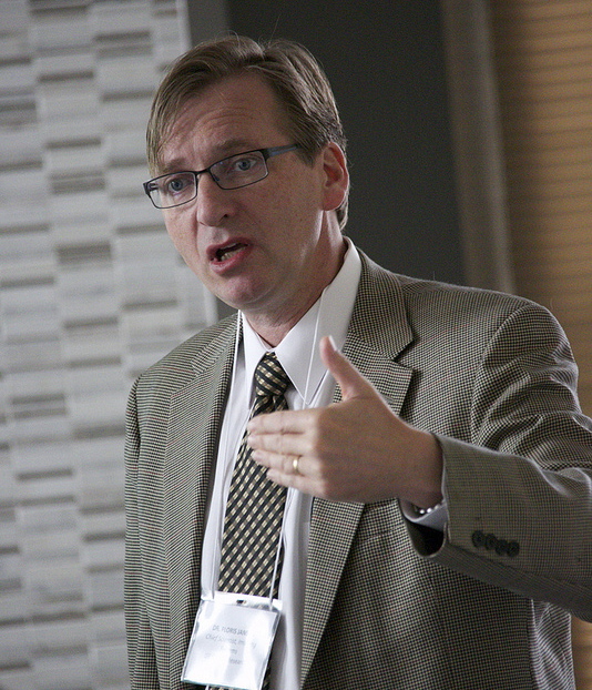
"When I think of hybrid imaging, I think of bringing other aspects of the information out and making it available for our doctors," said Dr. Floris Jansen, chief scientist of GE Global Research Center and another presenter at the workshop.
Jansen explained that the goal of hybrid imaging is to develop more personalized and focused care for patients. It also helps to make work easier for health care professionals so they can spend more time caring for patients and less time dealing with technology.
"Just because you can do it doesn't mean it's easy," said Jansen of new methods of hybrid imaging. "You don't want to create additional burdens on the patients, on the staff, on the doctors or the site. You have to somehow make that process easier."

The workshop's specific goals were to summarize current techniques of hybrid imaging, to identify challenges and develop a strategy for tackling these issues. The workshop further discussed the growth of the nuclear-based hybrid medical imaging industry as well as patient needs and demands.
The event was also the ideal venue for building new collaborations between researchers, industry and government.
Throughout the workshop, participants identified the top four priorities for future medical imaging research and training. These included funding, collaborations, evidence-based medicine and infrastructure development.
Researchers discussed these topics during breakout sessions. The group's recommendations will be used to draft a proposal for potential funding through government and industry programs.
The workshop's recommendations focus on researchers and industry working together toward a common goal. Industry members must inform researchers of their needs, and researchers must focus on addressing these needs through their work.
By working together, both industry representatives and researchers will have increased access to resources and funding. As well, including a diversity of people on the team allows for wider access to a greater spectrum of skills and knowledge.
A specific example developed by the team was to increase the use of university liaison offices between industry and research.
"The world isn't short on great ideas that have the potential to improve medical imaging technologies, but we first need agreement among partners about those issues that should be addressed first," said Dr. Paul Babyn, head of the U of S College of Medicine's Department of Medical Imaging.
"This workshop is taking us to the next level: it's stimulating network building, change through group consensus and collaboration."
Babyn also stressed the ongoing need for collaboration if hybrid medical imaging is to be a success.
"We are going to need new collaborators and members of our team — we will need radiochemistry and radiopharmacy," said Babyn in his closing comments.
Hosting the workshop in Saskatoon was no accident: the U of S and Saskatchewan are growing centres for the research and development of hybrid medical imaging and nuclear science. As well, the university has a unique variety of animal and human health care research occurring on campus.
"Saskatchewan has the opportunity to lead and make an important contribution to health care," said Dr. Baljit Singh, associate dean of research at the WCVM.

U of S resources include the Canadian Light Source — Canada's only synchrotron — where scientists collaborate on new diagnostic and treatment techniques for people and animals.
Another vital resource is a new cyclotron that will be operated by the Fedoruk Centre. The centre will produce medical isotopes for use in Saskatchewan's new PET-CT scanner at the Royal University Hospital. Saskatchewan is also rich in uranium — an element commonly used as a marker in medical imaging.
U of S researchers also have access to a variety of imaging technologies at the WCVM where animal models are used to investigate diseases that affect animals and people.
