
Step forward for toe tip necrosis
When I began my summer research work at the Western College of Veterinary Medicine (WCVM), the last thing I thought I'd be doing was acting as a cameraman at a photo shoot.
By Heather Sparkes
Instead of peering through a microscope, I spent a few months looking through the viewfinder of a Nikon CoolPix L810.
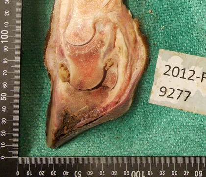
What's even stranger was my photo subject: cattle feet. Puzzle-piece sections of hoof, tendons and bones were part of the research that I was working on in an effort to better describe toe tip necrosis syndrome in feedlot cattle.
So how does a normal bovine foot acquire a lesion of toe tip necrosis?
The floor of the hoof (sole) attaches to the front of the hoof (dorsal wall) along what's called the white line. For some reason there is separation at this line, starting at the tip of the toe and working its way up the foot.
According to Dr. Chad Paetsch (WCVM '09), a case of toe tip necrosis is defined as separation along the apical white line and the presence of necrotic (dead) tissue in addition to lameness.
"The cases we have been seeing range from a necrotic lesion at the tip of the toe all the way to severe inflammation of the bones and tendons within the foot. In some cases infection has spread to the upper limb and/or lungs," says Paetsch, a WCVM graduate student who is studying the disease for his Master of Science program project.
No definite cause has been linked to the lesions, but it's believed that hyper-excitable cattle are at a greater risk of developing toe tip necrosis. The theory is these animals make their way through rough concrete handling facilities, paw the abrasive surface and wear down their hooves. That behaviour aids in white line separation and allows for bacteria to move from the environment into the foot.
Of the 59 cases received from feedlots across Alberta, the top three environmental contaminants cultured were Streptococcus, E. coli and Trueperella pyogenes. According to previous research, an inflammation with bovine viral diarrhea virus (BVDV) leads to vasculitis (infection of the blood vessels), both of which are risk factors for the development of toe tip necrosis syndrome.
Lameness in feedlot cattle has major implications for animal welfare and productivity. In the early 1990s, a study of five western feedlots found that lameness accounted for 16 per cent of animal treatments and five per cent of deaths. Of the lameness-causing pathology in the foot, three common types are foot rot, laminitis and toe tip necrosis syndrome.
Paetsch says the frequency of toe tip necrosis has increased since the 1990s — especially as practitioners are more accustomed to recognizing it.
"Because of the increased prevalence, I thought it would be interesting to provide more epidemiological data on this condition," says Paetsch, who is collaborating with his supervisor, Dr. Murray Jelinski, on the toe tip necrosis research.
To understand what makes toe tip necrosis cases different from healthy cattle, researchers collected feet from 148 euthanized animals at western Canadian feedlots. The feet were swabbed and cultured so researchers could test for bacterial and viral pathogens. Sections of claw were cut to test hardness, tensile strength, moisture and mineral content.
So far, the WCVM data shows that there is no seasonal or age predisposition for cases of toe tip necrosis versus lesion-free control animals. Affected cattle have been in the feedlot for a median of 27.5 days until they are euthanized. This is an early onset in comparison to laminitis that tends to occur at the end of the feeding period. Lesions may occur in all feet, but the outer claw of the hind foot is most often affected.
To provide the correct treatment, Paetsch says it's important for veterinarians to thoroughly examine the feet once they notice lameness. Instead of trimming the toe, partial amputation of the digit in the field is now recommended.
Supportive care, appropriate antimicrobial use, anti-inflammatory drugs and the placement of a wood block on the cow's opposite hoof claw are also suggested as ways to increase the likelihood of a successful treatment.
Once Paetsch finishes analyzing his project's research findings, he hopes to provide data that supports the efforts of practitioners and feedlot operators in reducing the future incidence of toe tip necrosis. Paetsch will present his research findings to western Canadian practitioners during the CanWest Conference that takes place in Banff, Alta., from October 19 to 22.
As for my picture taking, we analyzed my shots for thickness of the sole and dorsal wall as well as for measurement of angles to see if any coffin bone rotation exists along with this syndrome. I've taken about 600 images of cattle feet — enough images of toe tip necrosis to last my entire career.
Heather Sparkes of Arborg, Man., is a third-year veterinary student who participated in the WCVM's Undergraduate Summer Research and Leadership program in 2013.