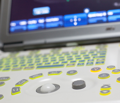
Ultrasound key to improving sample accuracy
A researcher at the Western College of Veterinary Medicine (WCVM) is conducting a study that will pinpoint the most effective and economical way to aspirate canine lymph nodes.
By Melissa Cavanagh
Lymph node aspiration is a diagnostic tool commonly performed by veterinarians. The technique involves using a thin needle to extract cells from a lymph node so they can be sent off to a lab and examined under a microscope.
While many veterinarians still manually aspirate lymph nodes, the key is using ultrasonography to increase accuracy and reduce costs for pet owners, says Dr. Monique Mayer, a veterinary radiation oncologist at the Western College of Veterinary Medicine (WCVM).
"We found that fairly often when veterinarians did an aspirate that wasn't ultrasound guided, they didn't get a sample that was useful — they didn't get any lymph node sample," says Mayer, who is also an associate professor in the college's Department of Small Animal Clinical Sciences.
With financial support from the WCVM's Companion Animal Health Fund, Mayer will study whether using ultrasound to guide the needle will increase accuracy of the procedure and the likelihood of obtaining a useful sample.
From her own experience, she says the ultrasound-guided technique allows veterinarians to see exactly what's happening: "You look with the ultrasound to see the node, and you also can see your needle coming in so it will guide your needle into the node."
Mayer adds that the increased use of ultrasound-guided lymph node aspirations could ultimately reduce costs for pet owners — thanks to fewer return visits and better lab samples.
Lymph nodes are located all throughout the body and serve as defence mechanisms. They help remove noxious agents such as bacteria and toxins, and they are the body's source of lymphocytes (white blood cells that are important for fighting disease).
When a lymph node swells, it indicates that the area being drained by that specific node is battling some sort of illness such as a bacterial infection or something more serious such as cancer.
"They [the lymph nodes] can get as big as a golf ball or even a tennis ball in certain disease conditions like lymphoma," says Mayer.
Aspirations are relatively non-invasive procedures that are conducted while a dog is awake. When veterinarians perform aspirations manually, they feel for the node and then insert the needle based on what they can feel.
"I'm usually doing aspirations to see if a tumour has spread to the lymph node because that will change how we treat the patient," says Mayer, explaining that information gained from aspirations is useful for staging a cancer.
"But they're also done for other reasons, non-cancer reasons – to look for signs of infection or other disease in the lymph node."
The problem is that not all diseased lymph nodes swell to a large enough size to be felt.
"Even when they have cancer they're not necessarily enlarged," says Mayer. "They've shown with oral melanoma (mouth cancer), for example, that the size of the lymph node didn't really predict whether there was cancer in it. A lot of normal-sized nodes did have cancer, so we need to test those."
Since lymph nodes can be difficult to feel, the needle often misses the node during an aspiration. And if the sample doesn't contain cells from the lymph node, veterinarians have to repeat the procedure — increasing costs for pet owners and forcing them to make multiple visits to their veterinary clinics.
Since many private veterinary practices already have ultrasound machines, this new approach is practical and a safe means of increasing aspiration efficacy, says Mayer.
"There haven't been any long-term risks associated with ultrasound wave exposure," says Mayer, adding that veterinarians may need to shave a small patch of the dog's fur around the site of the node.
Mayer plans to use canine patients at the WCVM Veterinary Medical Centre (VMC) as subjects for her study. She will perform both manual and ultrasound-guided aspirations of lymph nodes on dogs that are enrolled in the study so the viability of the samples can be compared.
Lymph nodes from the dogs' neck and "knee" region will be sampled, and only dogs without any history of lymph node disease will qualify for the study.
Mayer expects the project to be completed in one to two years.
"Getting clients whose dogs meet the criteria and are willing to agree to the study – it takes a while."
Melissa Cavanagh of Winnipeg, Man., is a second-year veterinary student and the WCVM's research communications intern for the summer of 2013.
