
Discovering the equine small intestine
Until recently, the inside – or lumen— of a live horse's small intestine was beyond the reach of traditional imaging modalities and remained a mystery to veterinarians.
By Joscelyn McKenzieBut a group of researchers at the University of Saskatchewan (U of S) have been working on a diagnostic tool that promises to overcome these limitations.
In collaboration with Khan Wahid from the U of S College of Engineering, Drs. Joe Bracamonte and Julia Montgomery from the Western College of Veterinary Medicine's (WCVM) Department of Large Animal Sciences have been testing the application of pill camera technologies and procedures that will enable them to capture valuable images of a live horse's small intestine.
In human medicine, pill cameras are used to diagnose small intestinal disorders such as Crohn's disease, polyps, tumours, and stomach ulcers. The U of S researchers are optimistic that their pill camera has the potential to effectively diagnose similar conditions in the horse and will also be valuable for checking surgical sites – small intestinal anastomoses, or connections – and providing information about the appearance and function of a healthy intestine.
Adapting the human set-up to work in horses was challenging for the researchers who encountered problems with getting the sensors to work consistently – particularly since a horse's abdomen is quite expansive when compared to that of a human.
The scientists began by placing the sensors as individual patches on the horse – the placement method used in humans. However, they had difficulty finding the optimal placement for each sensor that would ensure a consistent signal while the camera traveled through the many twists and bends of the horse's intestine.
They also discovered that their placement options were limited since each sensor was attached to the monitor by a wire that could only reach so far around the horse's belly. To overcome this obstacle, the engineering team used a newer sensor belt technology. The belt was a great advancement that provided a more consistent camera signal and saved a lot of time and guesswork with sensor placement.
The team was also challenged by the pill's limited battery life. Because each horse is different and gut motility depends on many factors, it was hard to estimate the length of time required for the camera to pass through the small intestine. To add to their dilemma, the pill sometimes sat in the stomach for a while before passing into the small intestine.
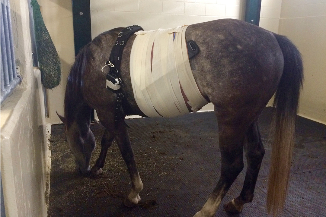
This initial pilot highlighted both potential applications and current limitations of the technology to be addressed. To begin the diagnostic procedure, the sensor belt is attached around the abdomen of the horse. Then the pill-sized camera is deposited into the horse's stomach using a nasograstric tube — a rubber tube that enters the horse's nose and extends down the esophagus into the stomach.
As the camera travels down the horse's gastrointestinal tract, it takes pictures at regular intervals and transmits a real-time signal which is picked up by the sensors attached to the outside of the horse's body. This signal is transferred from the sensors to a small monitor that's attached to a surcingle, a belt placed around the horse behind its shoulders. The images are then stored for later analysis and displayed so they can be seen in real time on the monitor.
Once the camera has left the stomach and entered the small intestine, the horse is fed hay to stimulate normal gut motility so the camera eventually exits the horse with the feces. The devices are single use only, so the researchers don't need to retrieve it later.
While the technology is showing great promise, there are several modifications required before it can be used clinically in horses.
Written by Joscelyn McKenzie. McKenzie, of Langley, B.C., is a fourth-year veterinary student who was part of the Western College of Veterinary Medicine's Undergraduate Summer Research and Leadership program last year. She was also the Townsend Equine Health Research Fund summer research student in 2016. Joscelyn's story is part of a series of stories written by WCVM summer research students.