
Angiostatin breathes new life into research targeting ARDS and cancer
It all began with the case of three kittens that were found in severe respiratory distress after a day spent in a laundry room with a home air purifier running.
By Kathy Fitzpatrick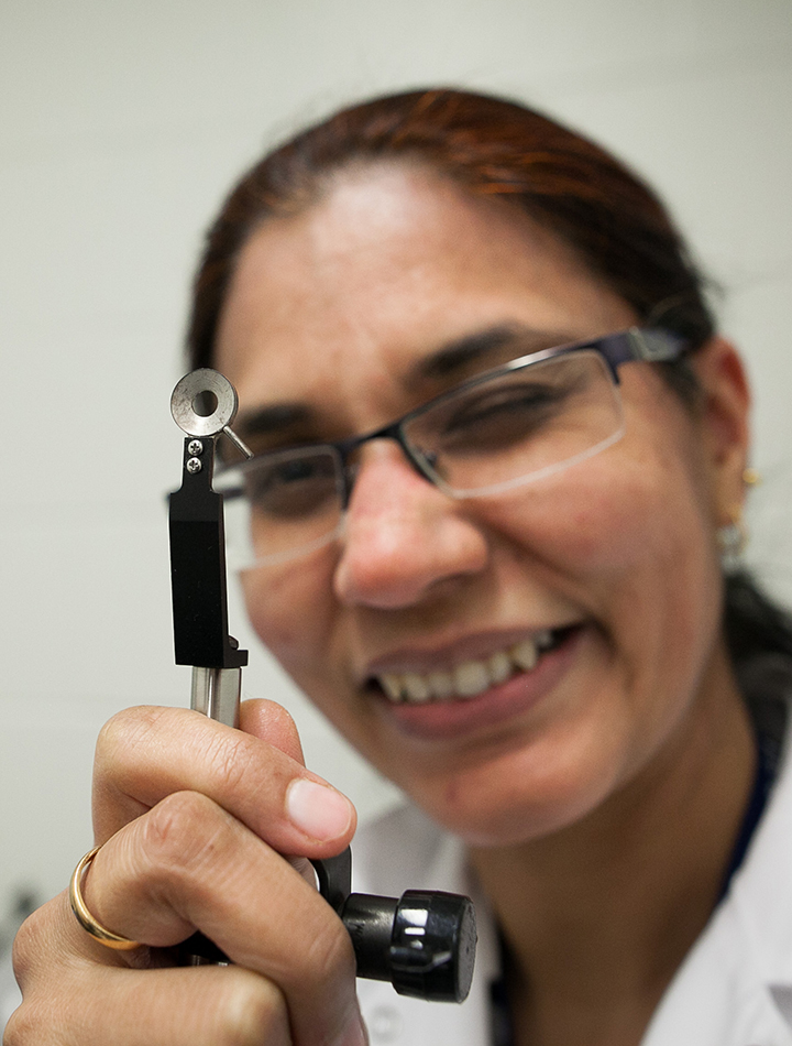
Ozone produced by the air purifier in the enclosed space had caused fluid to build up in the kittens’ lungs. The animals were suffering from acute respiratory distress syndrome (ARDS) — a potentially deadly condition in animals and people.
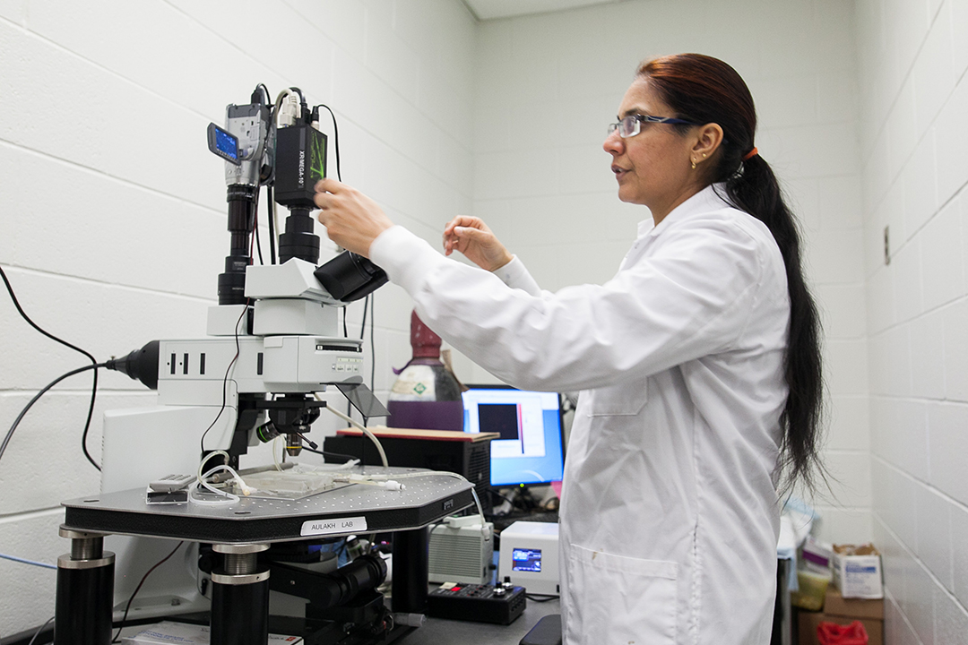
“Can you believe that a home air purifier in a room can produce ozone to concentrations that are believed to be harmless by the environment officials — but [are] not?” says Dr. Gurpreet Aulakh, who holds the Fedoruk Chair in Animal Imaging at the University of Saskatchewan (U of S).
Fortunately, once veterinarians placed the three young cats in a tent with enriched oxygen, the trio recovered. But their case has had a lasting effect on health research: Aulakh is now using animal models with ozone-damaged lungs as research models. Her hope is to find a way to diagnose ARDS at a much earlier stage than the current imaging technology permits. If she’s successful, her investigations could eventually have huge implications for both human and animal health.
ARDS develops when fluid builds up in the lungs’ air sacs following injury or infection. When patients can’t take in enough air, their organs are deprived of oxygen. Both physicians and veterinarians can use ventilation to treat their patients, but according to recent estimates, between one third and one half of patients diagnosed with ARDS die from the condition.
Of those who survive, some may have lasting lung damage.
In addition to developing better diagnostic tools, Aulakh’s research may also lead to new, more effective therapies for ARDS. Her attention has been drawn to two elements, one that may protect against lung damage and one that may actually cause damage.
Ironically, the latter is the neutrophil — one of the ‘foot soldiers’ of the immune system. Neutrophils are a type of white blood cell that are the first to arrive at the site of inflammation or infection and begin killing microbes.
However, neutrophils can also act like a body’s ‘frenemy’ — producing destructive chemicals that damage the structure of the lung.
Aulakh believes the key to stopping the damage is to prevent neutrophils from migrating into tissues. The element that may offer protection is angiostatin, a drug used to inhibit the growth of new blood vessels in malignant tumours. Aulakh has just presented some preliminary data at an imaging conference showing encouraging results.
Her team’s hypothesis is that complexes of angiostatin and related proteins protect against lung inflammation by preventing neutrophil migration into inflamed tissues. Through their research, Aulakh and her team hope to discover how exactly the complexes do this.
Aulakh is using imaging equipment at both the Canadian Light Source synchrotron and the Saskatchewan Centre for Cyclotron Sciences to test the hypothesis. Using a microscope, Aulakh follows the path of fluorescently-tagged angiostatin and its effect on immune cells in live mice with lungs damaged by ozone. She’s also looking at the extent and degree of lung damage.
In the next stage of her research, Aulakh will develop new biomarkers for positron emission tomography-computed tomography (PET-CT) imaging, which could aid in both diagnosis and treatment. Her final goal is to find a way to detect ARDS before symptoms appear. This may allow early intervention and treatment before the damage leads to death or irreversible damage.
Finding an effective way to diagnose and start treatment earlier for this disease could help save animals without the use of a ventilator would be the “holy grail,” says Dr. Elisabeth Snead, associate dean of research at the Western College of Veterinary Medicine (WCVM).
In veterinary health care, using a ventilator to treat animal patients diagnosed with ARDS is too expensive for most animal owners. Finding a more cost-effective treatment would allow veterinarians to give their clients another more viable option.
Snead is helping with the ozone model in Aulakh’s research. The associate dean was also instrumental in finding additional funding for Aulakh’s research program.
From the standpoint of pure science, Aulakh says studying ARDS is a quick and effective way of testing the basic biology of lung injury, which may shed more light on diseases affecting other organs.
As well, understanding more about lung injury could lead to significant advances in Western Canada’s livestock industry. Aulakh points out that cattle are “humongously affected by respiratory disease,” resulting in significant economic losses.
With her expertise in intravital microscopy and nuclear-based imaging, Aulakh is just the sort of person that Snead was looking for to join the WCVM’s Department of Small Animal Clinical Sciences and to fill the imaging research chair position. Snead wanted someone to help “ignite and excite” faculty and students about using radioisotopes for the diagnosis and treatment of various diseases in animals and people. She especially looks forward to new discoveries that successfully tag an antibody or drug onto an radioisotope for delivering treatment directly to the site.
Developing more expertise in this area is particularly important to the WCVM, given the expected addition of a large animal PET-CT scanner by January 2019.
Angiostatin also figures prominently in Aulakh’s second line of research: she’s focusing on an aggressive type of canine cancer called hemangiosarcoma that arises from the lining of blood vessels. Once dogs are diagnosed with this type of cancer, they generally live for only another six to eight months following surgery and chemotherapy.
Her goal is to find a better way to detect the disease at an earlier stage by investigating a protein called ATP-synthase. She and Dr. Humphrey Fonge, a clinical assistant professor in the U of S College of Medicine’s Department of Medical Imaging, are collaborating on the project.
As she points out, cancer appears to be even more prevalent in the veterinary world than on the human side — and that’s why she views her research as an opportunity to make unique discoveries that will benefit both animals and people. She’s particularly grateful for the chance to serve in a role that helps to bring together the university’s synchrotron and cyclotron, the veterinary college, the PET-CT imaging facility and the Royal University Hospital.
"I think these five entities are the enablers of some unique research, and I thank the Fedoruk Centre for this," Aulakh says. "They are probably bringing things together from all walks of science."
The Fedoruk Chair in Animal Imaging is supported by the Sylvia Fedoruk Canadian Centre for Nuclear Innovation Inc, a not-for-profit corporation funded in large part by Innovation Saskatchewan.