
New CT scanner creates more slices of life
Drs. Lesley Zwicker and Sally Sukut can’t hide their enthusiasm for the Western College of Veterinary Medicine’s new computed tomography (CT) scanner that’s pushing the boundaries of veterinary medical imaging.
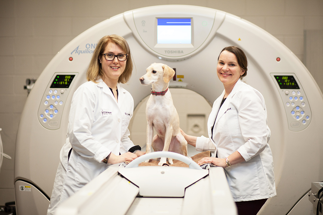
By Lynne GunvilleAs the board-certified radiology specialists explain, the Toshiba Aquilion ONE can create 320 “slices” or cross-sectional images during each rotation around the patient.
“With the 320-slice CT there are 320 detector rows, so we can scan a larger volume of the patient in a single revolution,” explains Zwicker, an assistant professor in the WCVM’s Department of Small Animal Clinical Sciences.
“We’re able to scan 16 centimetres of tissue in a half second, and that means we can acquire a whole region of the skull or brain or the heart, all in one rotation.”
The new unit replaces the college’s previous scanner that could produce 16 image slices per rotation. The Toshiba Aquilion ONE with its increased volume capabilities, advanced technology and improved software produces a clearer image with reduced artifacts — distortions or errors in the image that can make interpretation difficult.
The new CT scanner also allows members of the WCVM’s medical imaging team to reduce motion artifacts in regions where it has notoriously been an issue, such as the heart and lungs. The advanced software can produce images showing an organ’s structure, movement and blood flow in real time.
Dynamic imaging will also provide research opportunities such as perfusion studies involving the heart or brain. While both Zwicker and Sukut plan to incorporate the CT unit into their prospective research projects, they also point out that the machine can play a large role in recruiting other specialists to the WCVM.
For example, veterinary cardiologists and neurologists would welcome the opportunity to use such a high-tech CT machine for their clinical patients as well as for their research.
“The machine also offers unique research opportunities for our graduate students,” says Zwicker. “It makes sense for us to utilize this machine as much as we can to provide novel research opportunities for our residents and for the master’s or PhD students who are affiliated with the WCVM or even the University of Saskatchewan.”
WCVM veterinary students get a first-hand look at the advanced technology and learn more about its use as a tool for veterinary diagnostics and research studies. In addition, the CT scanner contributes to the WCVM’s goal of becoming a leader in cancer research and care.
“With our oncology patients, not only are we imaging the primary tumour but we’re also imaging the other regions of the body including the thorax and abdomen in order to stage them and monitor the appropriateness of treatment and regression of the tumour,” Sukut says. “The machine will allow us to acquire images a lot quicker when doing full body scans looking for metastases.”
Both Zwicker and Sukut look forward to providing exceptional diagnostic services for their clients as they investigate advanced clinical applications such as cardiac imaging and brain perfusions – applications that have already made significant contributions to human medicine.
“I think because the scanner’s dynamic capability is novel in veterinary medicine, it will impact the types of clinical services that we can offer as well as the research capabilities and collaborations,” says Zwicker.
“As a referral academic institute, it’s very important for the WCVM to stay on the cutting edge in the technology world with imaging. This new scanner allows us to be that sort of leader.”