
First cases show potential of PET-CT unit’s diagnostic capacity
The new technology has only been operating for two months, but Canada’s only PET-CT unit dedicated to clinical use in animals is already improving the care of patients at the Western College of Veterinary Medicine (WCVM).
By Jeanette NeufeldThe new technology has only been operating for two months, but Canada’s only PET-CT unit dedicated to clinical use in animals is already improving the care of patients at the Western College of Veterinary Medicine (WCVM).
The regional veterinary college celebrated the official opening of the Allard-Roozen Imaging Suite, which houses the new PET-CT unit, on June 7. The suite’s construction and purchase of the PET-CT scanner was made possible by a $2.5-million donation from Cathy Roozen, an Alberta businesswoman and philanthropist.
“It’s like being on the precipice of a big adventure from the point of view of being able to help our patients and just understand the mysteries behind the physiology and disease processes,” says Dr. Elisabeth Snead, associate dean of research at the WCVM.
When the PET-CT unit isn’t being used to help diagnose and treat clinical cases at the WCVM, University of Saskatchewan (USask) researchers will use the advanced technology for collaborative research studies that will benefit animals as well as people.
A PET-CT, or positron emission tomography-computed tomography scan, provides a diagnostic advantage over other imaging modalities by combining the detailed anatomic information from a CT scan with the comprehensive images of blood flow and metabolically-active structures attained with a PET scan.
This allows clinicians to measure changes in cell activity in the body and enables them to detect cancers, brain disorders and heart disease before anatomical changes are detectable by other imaging scans.
“It’s quite unique still. I think we are lucky to have one here,” says Dr. Kanae Takada, a resident in small animal internal medicine at the WCVM.
As part of this spring’s training process, clinicians and researchers offered PET-CT scans to two clients of the WCVM’s Veterinary Medical Centre (VMC). Having access to this highly advanced medical imaging has already proven beneficial for these first two patients.
A better end of life for a beloved cat
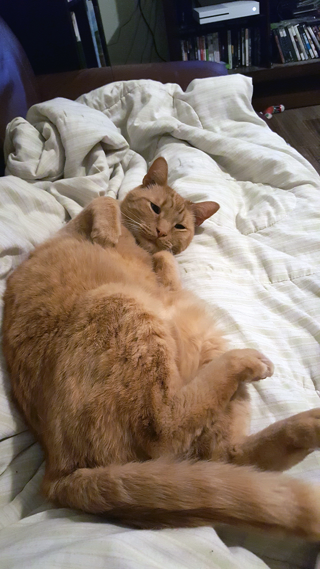
Jack isn’t like all cats – his owner Melissa Hylkema describes him as a “really, really friendly cat” who is generous with his affection.
Hylkema, whose family runs a dairy farm near Hague, Sask., was referred to the VMC after Jack began displaying strange neurological signs – he wasn’t eating or drinking and one pupil was dilated.
She chose to pursue a diagnosis because of Jack’s personality and her connection with the gentle, patient cat. She has owned him for his entire life, and the 11-year-old Jack loves to cuddle with Hylkema’s one-year-old daughter.
“I think any other cat wouldn’t be so receptive to spending that much time at the vet and going through these procedures,” she says.
After check-ups from the WCVM’s neurologist and ophthalmologists, the specialists narrowed it down to an issue with a nerve in his skull — but they were unable to tell if it was a tumour or another form of inflammation. Ultrasounds, X-rays and bloodwork came up inconclusive.
“Not knowing is always really challenging for the owner,” says Takada, who has overseen Jack’s case and recommended him for a PET-CT scan.
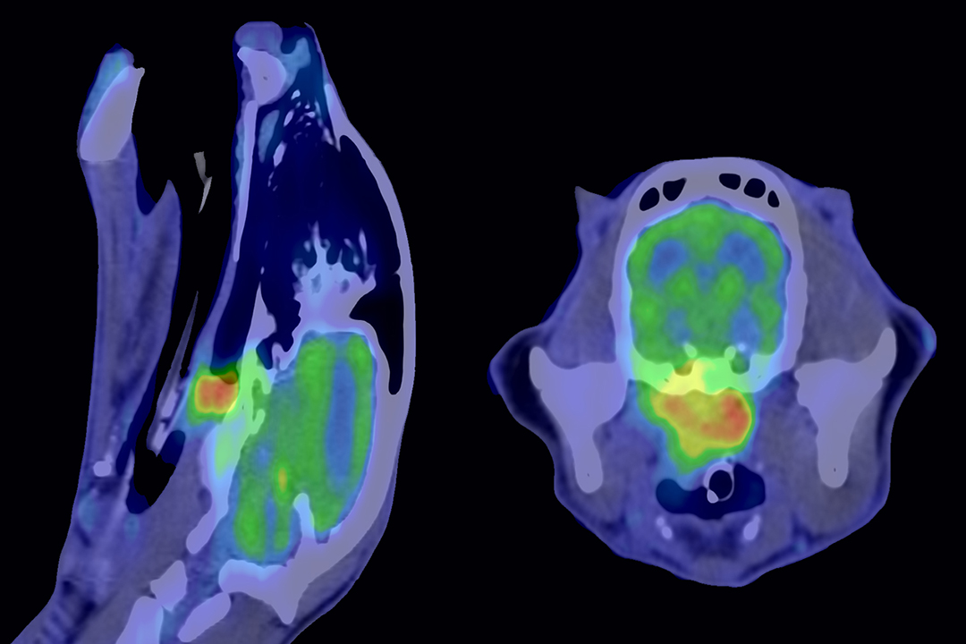
By imaging Jack’s head and neck using the PET CT, the clinicians could see a mass in the cat’s nasal pharyngeal area and detect that cancerous cells had spread to one of his local lymph nodes. They then took a cell sample using a small needleto determine the cancer diagnosis.
While the diagnosis wasn’t positive, Jack’s owners are able to better understand his disease and offer him palliative care to ease the end of his life.
“It helps that I wasn’t sitting around wondering if there’s something else I could have done,” says Hylkema.
Solving the mystery behind a recurring infection
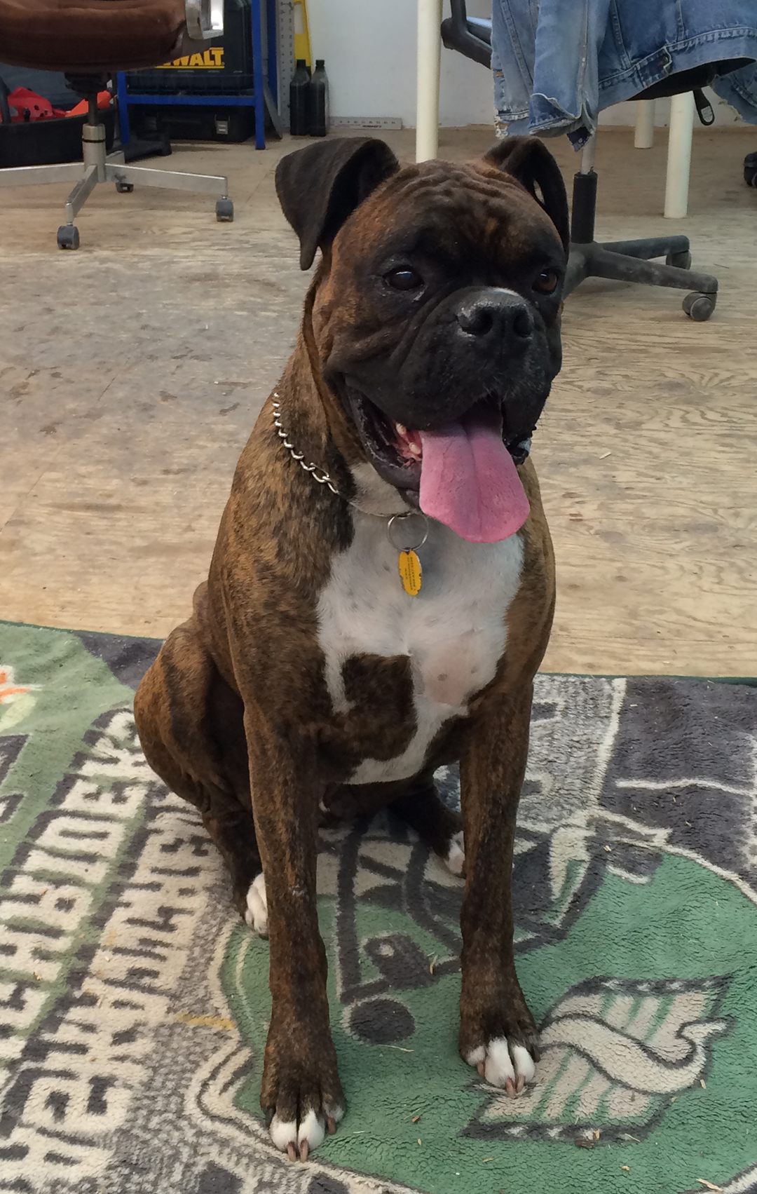
Levi, a four-year-old boxer, has suffered from chronic infections since he was a puppy, and despite continued rounds of antibiotic drugs, these infections would never fully clear up. His owner, Mark Forsyth, describes Levi’s symptoms as severe pain in his hind end.
“He could hardly sit down,” he says. Once, his right leg filled up, as if with water. “If you squeezed it, it was like a wet sponge.”
Forsyth, who lives in Oxbow, Sask., was referred to the WCVM by his local veterinarian, Dr. Marcel MacFarlane at Souris Valley Veterinary Services.
By using the advanced PET-CT imaging, WCVM clinicians located the source of infection and confirmed MacFarlane’s suspected diagnosis.
Levi was suffering from a condition called discospondylitis, a deep bacterial infection in the spine. The disease can cause severe pain as well as nerve damage in dogs if left untreated.
“He was in severe pain. A delay in the diagnosis could have delayed initiation of treatment,” says Takada.
She explains that without the PET-CT scan, the WCVM team would have had to rely on multiple tests and other forms of medical imaging, potentially taking more time and subjecting Levi to more than one round of general anesthesia.
“Because he already had lots of chronic change, [it] would have been confusing if we didn’t go straight ahead to the PET-CT. We may have had extra steps to reach the diagnosis,” she says.
Levi’s clinical signs were also unusual, which added to the challenge of diagnosing his condition.
After making the diagnosis, clinicians sent Levi home, and he is now recovering on an extended course of antibiotics.
“He’s doing good,” says Forsyth, who describes his dog as “laid back” despite his continued health challenges.
More than a month after his PET-CT scan, Levi remains symptom-free and his owner is relieved to have a diagnosis.