Putting shock wave therapy to the test
Dr. Angela MacKay’s passion is solving equine lameness.
By Katie Brickman-Young“My favourite thing to do is try to sort out where the horse is hurting and how to make it feel better,” says MacKay, a clinical associate in equine field service at the Western College of Veterinary Medicine (WCVM).
When MacKay was completing her combined residency and graduate program at the WCVM, her graduate research project focus was on the use of an alternative therapy — extracorporeal shock wave therapy (ESWT) — to treat caudal heel pain.
Her thesis topic was based on a suggestion from her supervisor, WCVM assistant professor Dr. Kate Robinson, whose own horse was affected by caudal heel pain. The desire to look at ESWT was due to the lack of research around the use of a therapy that’s commonly used to treat caudal heel pain in horses.
“People are prone to jump on the bandwagon for alternative therapies without understanding what is happening and if what they are doing is helpful,” says MacKay. “That’s why I thought it was a great idea to study shock wave therapy and its effects.”
A ‘perfect storm of events’
Caudal heel pain is a catch-all term used to describe pain in the heel region of the foot, usually in the horse’s front limbs, explains MacKay. Most owners know caudal heel pain as navicular syndrome — pain coming from the navicular region of the foot.
In addition to the navicular bone, this vital area consists of soft tissues, tendons and ligaments, as well as the coffin joint and fluid-filled structures that assist movement.
“The navicular region is important for supporting the foot but also for allowing the coffin joint to flex,” explains MacKay. “All the structures can be injured when navicular syndrome or caudal heel pain occurs.”
Veterinarians believe that 90 per cent of lameness in horses is within the foot, but from that percentage, it’s unclear how many lameness issues are considered navicular syndrome. Researchers think caudal heel pain is a “perfect storm of events,” a degenerative condition that results in an abnormality of one or more of the equine foot’s structures.
“In normal tissues in any animal or human, there are micro-injuries when we use our bodies. Usually those heal and make tissues stronger over time. In the foot of the horse, the blood supply is different, and there is the thought that these structures get those micro-injuries, but they can’t heal normally for some reason,” says MacKay.
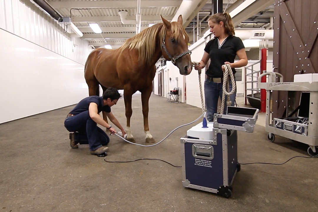
In shock wave therapy, the machine generates a sudden energy of release to cause a pressure wave, which can stimulate the cells by changing their membranes — encouraging them to release growth factors and increase cell metabolism.
“It feels like a hammer hitting the tissues. It does cause a bit of discomfort when applied, so we sedate the horses before treatment,” says MacKay. “Shock wave therapy can help, potentially, by having some anti-inflammatory effects and increase blood supply to the region.”
Two-phase study
In the first phase of their research study, MacKay and Robinson surveyed members of the American Association of Equine Practitioners through an online questionnaire about the condition. Of the 144 veterinarians who responded, 65 practitioners frequently used shock wave therapy to try and treat ligament-related injuries.
In the study’s second phase, the research team put a video together asking for local horse owners who would be interested in participating in the study. After receiving 200 requests, they evaluated 83 horses and worked with 30 client-owned horses that were all diagnosed with caudal heel pain in one or more of their limbs.
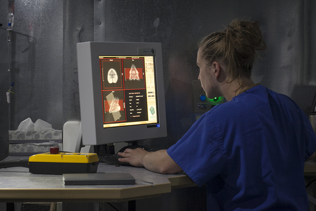
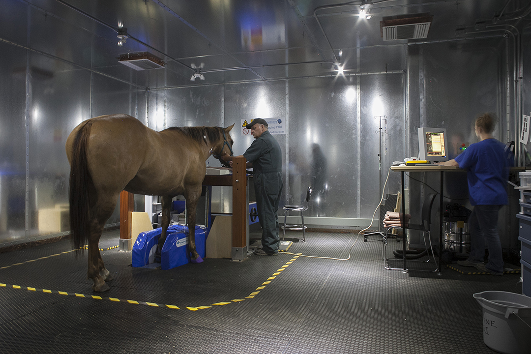
For each horse, MacKay and Robinson completed a lameness exam and physical exam, nerve blocks, X-rays of the front limbs (if a horse passed the nerve block step), and magnetic resonance imaging (MRI) of the affected limbs.
Then, the researchers randomly slotted each horse into one of two groups. Horses in the treatment group were sedated and received shock wave therapy every two weeks for a total of three treatments, while horses in the control group were sedated but received no treatment over the same period.
Throughout the study, the researchers conducted lameness exams and kinematic gait analyses on all horses before sedation and treatment. At day 128 (32 weeks after the initial treatment), the research team repeated all of their original evaluation steps.
Based on the study’s results (see below), MacKay and Robinson concluded that ESWT was not a supportive treatment option for navicular syndrome or caudal heel pain.
“Horses simply did not experience improvement, for whatever reason,” says MacKay. “As with any research project, there are limitations that may have affected our results, but I don’t recommend shock wave therapy for navicular syndrome because I think there are better options.”
Financial support for this study came from the Mark and Pat DuMont Equine Orthopedics Research Fund, Saskatchewan Agriculture Development Fund, and the Townsend Equine Health Research Fund. TEHRF also provided scholarship funding.
Click here to read more stories in the Summer 2020 issue of Horse Health Lines, publication for the Townsend Equine Health Research Fund.
What we learned
After analyzing all of the study's data, here is a round up of the WCVM equine research team's key findings about the use of extracorporeal shock wave therapy (ESWT).
- Horses in the shock wave treatment group that had a single leg forelimb lameness experienced a statistically significant improvement in their lameness (pain) following treatment, based on subjective evaluation.
- Horses in the treatment group with a bilateral forelimb lameness (both front legs were in pain) did not experience a statistically significant improvement in their pain following treatment, based on subjective evaluation.
- Horses with a bilateral forelimb lameness can be difficult to assess accurately. The way they move to compensate for their pain sometimes inhibits the ability to get a real sense of their pain.
- All horses enrolled in the study had injury to more than one structure in the foot of the most painful leg. Most horses had an injury to their deep digital flexor tendon, had osseous fluid in their navicular bone (which can indicate edema or swelling, injury from repetitive concussion or degeneration), and had inflammation in their navicular bursa (sac cushioning the navicular bone from the deep digital flexor tendon). This condition is referred to as “navicular bursitis.”
Click here to read Dr. Angela MacKay’s thesis. Read "MacKay driven by helping horses," which profiles Dr. MacKay's academic career.