Chronic wasting disease (CWD) has intrigued the scientific community ever since its detection in a Colorado facility 44 years ago.
At the beginning, researchers attributed the clinical signs to stress, malnutrition or poisoning. Twelve years later, it became part of the class of prion diseases, and the only one known to infect both free-ranging and captive deer – to make matters worse, with a 100 per cent fatality!
As infective prions accumulate in the brain, they cause clear neurological signs, such as low head position, wide leg stance, decreased reactiveness and gradual weight loss. These deer usually die from starvation and aspiration pneumonia.
When I first arrived in Saskatoon, Sask., I had little idea of the severity of the situation. In the last three years, I have seen the CWD prevalence of studied deer grow from six per cent to 33 per cent in our CWD-endemic study area along the South Saskatchewan river valley.
Researchers here make enormous efforts to understand this spread and to promote responsible management practices and policies. It honestly took me no more than five minutes to fully appreciate this working group. Together, we seek to understand how movement patterns, use of congregation areas, and social structure of the mule deer herds affect the spread of the disease.
More specifically, at the University of Saskatchewan, Dr. Trent Bollinger and I investigate how different sex and age classes of mule deer use environmental sites. We know that prions deposited into the environment are persistent and continue to infect susceptible cervids for years. However, to date no one has managed to quantify the amount of prions in the environment and to determine the infectious dose that makes deer sick in the wild.
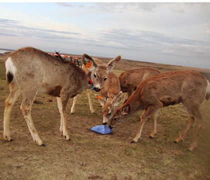
So, first, we need to untangle how and when deer use environmental sites in a CWD-endemic area. For that, we have been placing triggered-by-movement cameras in different locations since July 2009, including locations next to CWD-infected mule deer carcasses.
We performed a preliminary analysis to explore the potential and limitations of the data and the results are exciting. We found that carcasses are visited the least often and significantly less frequently than natural trails and rub sites.
Also, adult and juvenile males use anthropogenic sources of food (salt licks for cattle and sites where grain has spilled) most frequently. We think that if these sites are important sources of prions, our findings might partially explain the higher prevalence of CWD in males in our study area and reported elsewhere.
I'm both satisfied and optimistic about this first step into a behind the scenes look into the lives of mule deer. What's more, by incorporating all the available data we will be able to make better conclusions about how different age and sex classes of mule deer use different environmental sites.
These conclusions will enhance our understanding of the transmission dynamics and prevalence trends, which will help us to build better management strategies for controlling CWD in populations of wild deer.
Dr. Maria Fernanda Mejia-Salazar is a PhD student in the WCVM's Department of Veterinary Pathology. Originally from Mexico City, Mejia-Salazar recently received a prestigious scholarship from her home country. Mejia-Salazar's article was first published in the August-November 2011 issue of PrioNews. Reprinted with permission from PrioNet Canada.
At the beginning, researchers attributed the clinical signs to stress, malnutrition or poisoning. Twelve years later, it became part of the class of prion diseases, and the only one known to infect both free-ranging and captive deer – to make matters worse, with a 100 per cent fatality!
As infective prions accumulate in the brain, they cause clear neurological signs, such as low head position, wide leg stance, decreased reactiveness and gradual weight loss. These deer usually die from starvation and aspiration pneumonia.
When I first arrived in Saskatoon, Sask., I had little idea of the severity of the situation. In the last three years, I have seen the CWD prevalence of studied deer grow from six per cent to 33 per cent in our CWD-endemic study area along the South Saskatchewan river valley.
Researchers here make enormous efforts to understand this spread and to promote responsible management practices and policies. It honestly took me no more than five minutes to fully appreciate this working group. Together, we seek to understand how movement patterns, use of congregation areas, and social structure of the mule deer herds affect the spread of the disease.
More specifically, at the University of Saskatchewan, Dr. Trent Bollinger and I investigate how different sex and age classes of mule deer use environmental sites. We know that prions deposited into the environment are persistent and continue to infect susceptible cervids for years. However, to date no one has managed to quantify the amount of prions in the environment and to determine the infectious dose that makes deer sick in the wild.
So, first, we need to untangle how and when deer use environmental sites in a CWD-endemic area. For that, we have been placing triggered-by-movement cameras in different locations since July 2009, including locations next to CWD-infected mule deer carcasses.
We performed a preliminary analysis to explore the potential and limitations of the data and the results are exciting. We found that carcasses are visited the least often and significantly less frequently than natural trails and rub sites.
Also, adult and juvenile males use anthropogenic sources of food (salt licks for cattle and sites where grain has spilled) most frequently. We think that if these sites are important sources of prions, our findings might partially explain the higher prevalence of CWD in males in our study area and reported elsewhere.
I'm both satisfied and optimistic about this first step into a behind the scenes look into the lives of mule deer. What's more, by incorporating all the available data we will be able to make better conclusions about how different age and sex classes of mule deer use different environmental sites.
These conclusions will enhance our understanding of the transmission dynamics and prevalence trends, which will help us to build better management strategies for controlling CWD in populations of wild deer.
Dr. Maria Fernanda Mejia-Salazar is a PhD student in the WCVM's Department of Veterinary Pathology. Originally from Mexico City, Mejia-Salazar recently received a prestigious scholarship from her home country. Mejia-Salazar's article was first published in the August-November 2011 issue of PrioNews. Reprinted with permission from PrioNet Canada.
Share this story